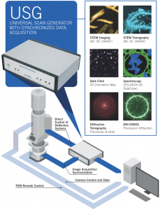
TVIPS has launched its newly developed Universal Scan Generator (USG) which allows to control up to eight TEM deflection coils or lenses with user-defined parameters directly in combination with synchronized image acquisition. The USG features 8 analog input/output channels (±10 Volt, 16 bit) and 64 digital input/output channels.
Thanks to its universal design the USG device is suitable for different applications:
Most TEM types can be retro-fitted with the USG device. In case of a TEM without internal scan generator, beam and image shifts are performed by controlling the deflection coils directly (above and below the specimen).
- Simultaneous acquisition of BF, DF, HAADF images (single or continuous mode)
- Up to 16k × 16k pixels image sizes
- Recording of individual Ronchigrams
- Input for camera synchronization signal
- AC synchronization
- Powerful image acquisition and processing software
See also EM-Scan for more information!
- Dynamic focusing
- Linear contrast: The Bragg contribution of crystalline specimen can be reduced
- Increased focal depth by means of nearly parallel illumination: Specimens up to 1 micron thickness can be studied (± 70 degree)
- Low dose tomography using a navigator tool
- Less radiation damage compared to conventional TEM
- Reduced dynamical effects (close to kinematical theory)
- Increased number of reflections in reciprocal space compared to conventional diffraction
- 3D Pivoting Diffraction: Leading to recording intensities of the whole reciprocal space of a single crystal under low dose conditions, e.g. for protein crystallography
- Powerful automatic image acquisition and processing software
- Automatic calibration procedure
See also EM-Conos for more information!
As a recently developed non-destructive technique*, the DFOM is utilized for 3D grain-oriented mapping in polycrystalline specimens with a spatial resolution down to 1nm. Thereby, a series of conical-scanning dark field images are acquired as function of stage tilt automatically. Typical settings parameter are:
- Stage tilt range: ± 60 degree, 1 degree incr.
- Beam tilt range: 360 images per ring, 10 rings
- Up to 400.000 images taken in a few hours
See also EM-DFOM for more information!

