Pivoting Diffraction Tomography (TEM)
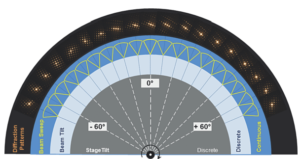 Automatic 3D Electron Diffraction Tomography (EDT) acquires full reciprocal data sets of single crystals under low dose condition such as organic or protein crystals (life sciences) and beam sensitive materials (material sciences). The combination of discrete stage tilt and discrete/continuous beam tilt (perpendicular to stage tilt axis) makes sure that diffraction reflexes of the whole reciprocal space of a crystal can be recorded using a minimum electron dose.
- A powerful acquisition and processing software for reconstruction
- Semi-automated calibration procedure
| Example for data acquisition with a total electron dose of about 8e/Ų | |
|---|---|
| Goniometer tilt range | ±30° |
| Goniometer tilt increment | 4° |
| Discrete beam tilt step | 0.2° |
| Beam tilt sweeping range | 0.2° |
| Exposure time | 500ms |
| Number of diffraction patterns | 336 |
| Dose per image | 0.024e/Ų |
| Total acquisition time | 8 min |
For this application the TVIPS Universal Scan Generator (USG) is required.
Precession Diffraction Tomography (S/TEM)

- The electron precession1 can be realized by beam tilt in a hollow cone using the four beam shift coils controlled by USG (up to 120 Hz). To compensate the diffraction pattern movement caused by precession the four image shift coils are simultaneously used.
- This technique facilitates high quality/ high resolution diffraction patterns from a zone axis due to reducing of the dynamical effects (close to kinematical theory).
- A perfect alignment of the crystal is unnecessary.
For this application the TVIPS Universal Scan Generator (USG) is required.
1 P. A. Midgley and A. S. Eggeman, Precession electron diffraction-Topical Review, IUCrJ (2015). 2, 126-136.