Navigation Software for Low-Dose Applications
This module was specifically developed for low-dose applications, in order to find the area of interest and to avoid spare exposures during searching or focusing. The overview images at lower magnifications are used to navigate and to define the positions at higher magnifications. This makes microscopy sessions more efficient because unnecessary TEM operations and exposures are avoided. EM-Navigation is supporting almost all TEM models (including in-column energy filter) and has been tested and installed on a wide range of TEMs, e.g. , e.g. FEI Tecnai12/20/30, Polara, Titan Krios, Hitachi H-9500, JEOL JEM-1230/1400/2010/2100/2200/3100/3200FSC, Philips CM100/120/200/300 and Zeiss 912/922, Libra 120/200, Kronos.
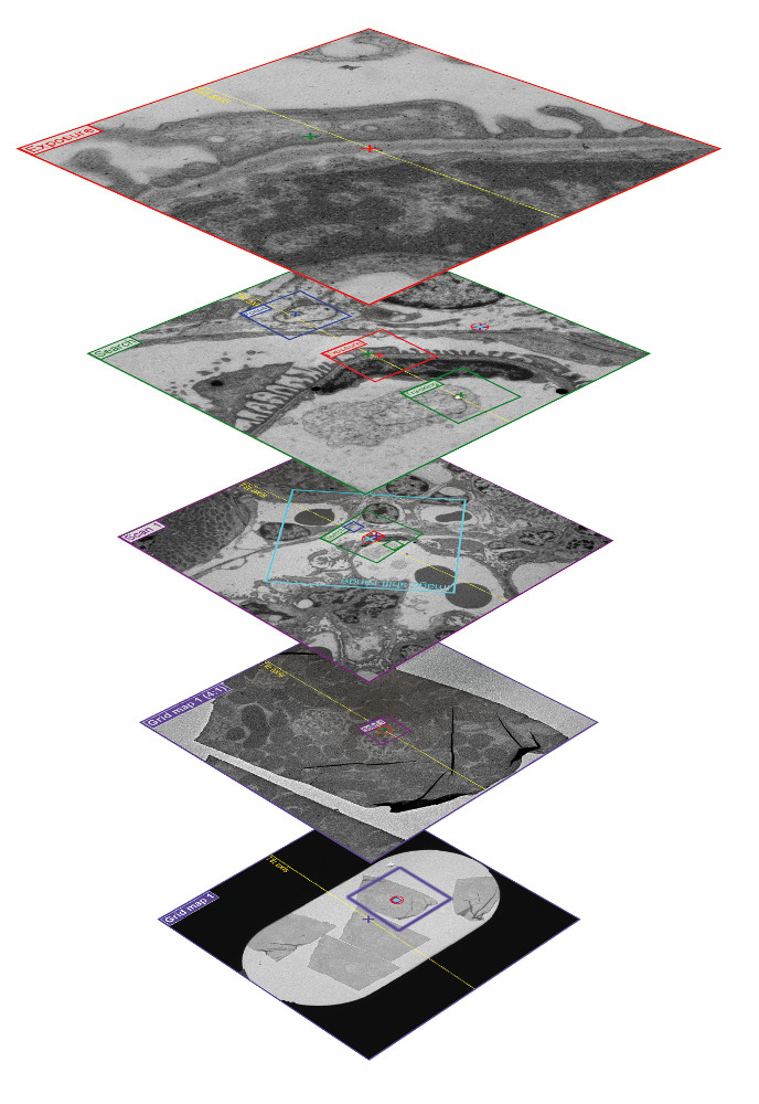
Grid map
First a large scale image (~ 1.5 x 1.5 mm²) is recorded at low magnification e.g. 120x. This image is composed of e.g. 5 x 5 tiles (20 480 x 20 480 pixel) and provides an overview of the grid and is used to screen throughout the sample without unnecessarily exposing the specimen.
Scan
At higher magnification (e.g. 1 100x with objective lens on) several areas of interest can be localized more precisely. It is possible to define multiple scan positions.
Search
Based on the previously defined scan positions the search position shows the area of interest at higher magnification (e.g. 4 400x). Typically on this position the eucentric height will be corrected automatically (auto-eucentric).
Focus
This position uses the same magnification like exposure position but is shifted e.g. along the tilt axis for the tomographic setup. On this position the defocus will be corrected automatically (auto-focus). For avoiding illumination of adjacent areas the beam size and position is centered automatically (auto-beam-centering).
Tracking
This position is mainly used for the tomographic setup and is shifted along the tilt axis of the stage. The tracking position is used to re-center the object for subsequent acquisitions.
Exposure
This position records the final image of the area of interest.
- Built-in navigation function for low-dose applications
- Compensation for displacements of different magnifications
- Automated focus adjustment
- Automated beam centering
- Automated eucentric height correction
- Drift measurement
- Position recentring
- Spotscan acquisition
- Batch tomography